
Back in June 2021 we reported on a study by a research group in Austria showing that endothelial dysfunction (layer of cells lining blood vessels) is another complex piece in the complex ME/CFS jigsaw (Scherbakov et al., 2020; Sørland et al., 2021; Blauensteiner et al. 2021).
What are the findings of this new study?
This research group (led by Dr Francisco Westermeier) has now expanded their study further showing that plasma from ME/CFS patients induces endothelial dysfunction in vitro (experimental work performed outside of a living organism, such as in a test tube) with their results being published in the Journal of Vascular Pharmacology.
This new study showed a decreased production of nitric oxide by the endothelium cells when exposed to plasma from people with ME/CFS, using samples from the UK ME/CFS Biobank (UKMEB) (see below).
Nitric oxide (NO) is needed for regulating blood flow throughout the body. Endothelial cells actively regulate the immune system, and NO helps to regulate blood and oxygen supply throughout the body.
The study suggests the enzyme involved in the endothelial dysfunction (nitric oxide synthase, eNOS) may be involved in the pathophysiology of ME/CFS.
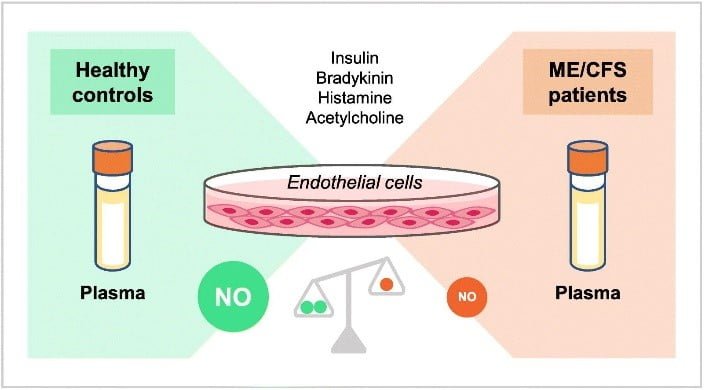
Graphic taken from the paper- showing the imbalance in NO in ME/CFS patients versus healthy controls.
In this research summary, with help from the authors, we have put these findings into context and help explain what these mean to people with ME/CFS.
What do the authors say about their findings?
Dr Francisco Westermeier said:
“We are (positively) surprised by these findings. This study represents one step forward because it is in line with our previous one, which addressed endothelial dysfunction-related markers in plasma from ME/CFS patients (Blauensteiner et al., 2021).”
How might NO production be involved in symptoms experienced by people with ME/CFS?
Nitric oxide (NO) gas is produced by endothelial cells lining the blood vessels and is known to promote vasodilation. This key vascular process allows oxygenated blood to reach our tissues all over the body. It is well-known that NO production increases in response to physical activity in healthy individuals.
However, ME/CFS patients might suffer from defective vascular function following exercise (as shown by Bond et al., 2021). Since post-exertional malaise (PEM) refers to a worsening of ME/CFS-related symptoms after minimal physical exertion, NO might certainly play a role in PEM.
Besides its crucial role in the vascular system, NO also serves as a versatile signal molecule at the immune and metabolic levels. Thus, although our findings in vitro (in a test tube) showed that plasma from ME/CFS patients reduced the ability of endothelium cells to produce NO, whether this highly reactive gas is involved in the debilitating multisystem-related symptoms should be also considered in further studies.
What could these findings of reduced NO mean for finding treatment options?
To the authors knowledge, these findings provide the first cellular evidence of impaired NO production in ME/CFS at the vascular level. A potential treatment could be pharmacological enzyme involved in producing NO, which is an endothelial isoform of nitric oxide synthase (eNOS). The activity of this enzyme was shown to be reduced in presence of plasma from ME/CFS patients.
Nevertheless, the results should be interpreted with caution, due to NO is a signal molecule produced as a response to multiple stimuli. An interesting example to better understand how this signal transduction is carried out is by comparing NO with an amplifier. A radio station has an antenna that emits radio waves (external signal), a radio (transducer) increases the strength of the signal (amplifier) to finally convert the original radio waves into sound waves (response). Our findings explored a simplified system involving a radio station (plasma from ME/CFS patients), a radio (endothelial cells, ECs), and an amplifier (NO).
Interestingly, listening to music is possible by using different devices (smartphones, conventional radios, TV, among others). In our body, there are different types of endothelial cells that produce (emit) NO (amplifier) with different patterns.
Our study used human umbilical vein endothelial cells (HUVECs) which have been extensively used in vascular biology research. Additionally, the listeners of the sound waves are vascular smooth muscle cells (VSMCs) which upon receiving the amplifier (NO) they increase the diameter of the vessels promote vasodilation (response). Although our findings did not evaluate the association among the radio (ECs), the sound waves (NO) and the listener (VSMCs) further studies involving this proposed experimental design might provide new pharmacological for treating endothelial dysfunction in ME/CFS.
Is this research group planning another study on endothelial cells?
The research group is interested in exploring whether the defective eNOS-dependent NO levels are correlated with increased reactive oxygen species (ROS) production in endothelial cells exposed to plasma from ME/CFS patients. The research group has based their assumptions on the fact that ROS leads to endothelial dysfunction by reducing the vascular bioactivity of NO in several diseases.
What do the researchers plan to do next?
Over the next months, the researchers aim to expand the existing evidence of endothelial dysfunction in ME/CFS by analysing several biochemical pathways and metabolites responsible for NO production. This new project is supported by Solve ME/CFS Initiative (USA) and carried out using samples from the UK ME/CFS Biobank (UKMEB).
Use of the UK ME/CFS Biobank
This study was funded by ME Research UK and used 29 samples from healthy controls and 58 ME/CFS samples from the UK ME/CFS Biobank (UKMEB).
The UKMEB was launched in August 2011 with funding and input from the ME Association (MEA), Action for M.E. and ME Research UK. The MEA Ramsay Research Fund has continued to provide annual grants since 2011 and now covers all the basic running costs of this vital and unique project.
The UKMEB contains samples from mild to severely affected people aged 18-60, as well as healthy controls and people with multiple sclerosis (MS). Blood samples have been aliquoted into serum, plasma, peripheral blood mononuclear cells (PBMC), red blood cells/granulocyte pellet, whole blood, and RNA. They use the samples in their own research and provide them to research teams from around the world. More information on this ME Association funded project can be found here.
We asked Dr Francisco Westermeier why he chose to use the UKMEB for obtaining samples for his study. He said:
“Late 2018 our research group received an ‘award to enable the study of human tissue samples / clinical data for biomedical research into ME/CFS’. This excellent opportunity – I saw posted on Twitter – along with the financial support from ME Research UK (MERUK) were crucial to initiate our research line in ME/CFS.
We are also grateful to patients for donating their blood to UKMEB, the ME/CFS community who raised funds to facilitate the UKMEB projects and the ME/CFS Society Austria (Österreichische Gesellschaft für ME/CFS, CFS-Hilfe) for supporting our research grant applications.”
Acronyms used in this summary
| Endothelium cells | EC |
| Human umbilical vein endothelial cells | HUVEC |
| Multiple sclerosis | MS |
| Nitric oxide synthase | eNOS |
| Nitric Oxide | NO |
| Peripheral blood mononuclear cells | PBMC |
| post-exertional malaise | PEM |
| Reactive oxygen species | ROS |
| Vascular smooth muscle cells | VSMC |
| UKMEB | UKMEB |
We would like to thank Dr Francisco Westermeier for helping us write this summary and answering our questions.
Dr Katrina Pears,
Research Correspondent
The ME Association
