Dr Krista Clarke is a post-doctoral researcher at the University of Surrey, and is currently working on a research study, co-funded by ME Research UK and the ME Association, assessing the electrical properties of white blood cells in ME/CFS. In this article, Krista talks about her PhD work on the electrophysiological properties of cells, and how this has led on to her research in ME/CFS.
Electrophysiological Properties of Cells in Health and Disease
During my PhD at the University of Surrey, the overall aim of my thesis was to investigate how the electrophysiological properties of cells change in health and disease as biomarkers for biomedical applications.
My PhD supervisors included Professor Mike Hughes, Dr Fatima Labeed, Dr Rebecca Lewis, and Professor Rob Dorey. All work on ME/CFS was also in collaboration with Mrs Caroline Kingdon and Dr Eliana Lacerda at the London School of Hygiene and Tropical Medicine.
The Electrophysiological Properties of Cells
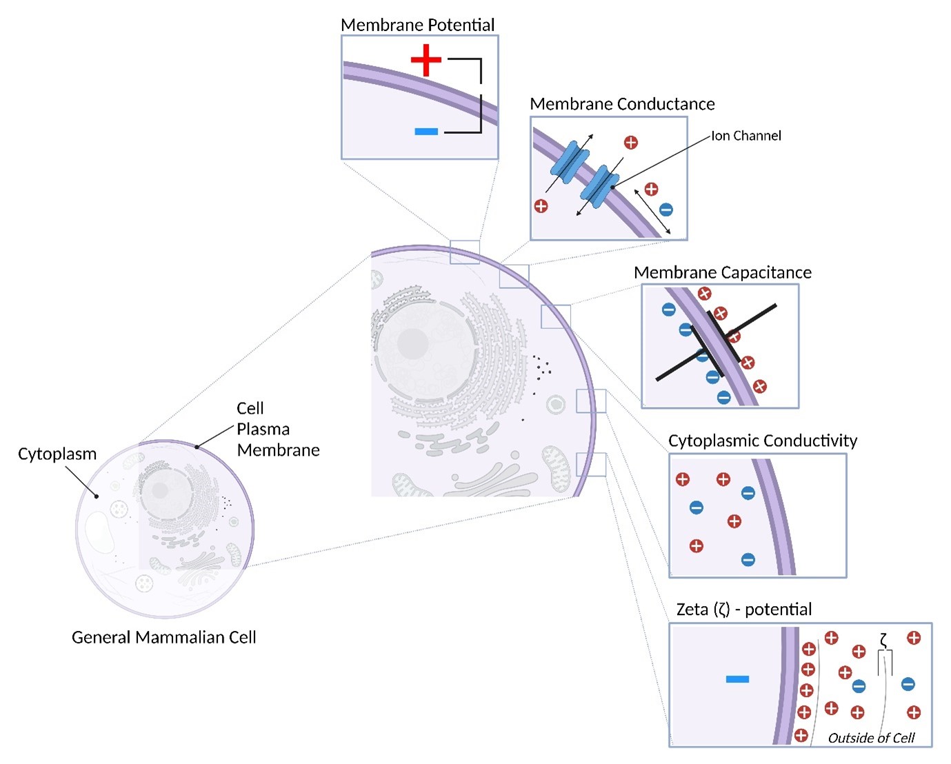
As seen in the diagram below, all mammalian cells consist of a plasma membrane surrounding a gelatinous liquid called the cytoplasm, composed of water, salts, ions, and other compounds. The plasma membrane is made of lipids, and acts not only to hold the cell together, but also as a barrier to tightly control and regulate which molecules and elements can enter and leave the cell – to control the cell’s internal environment.
Positively or negatively charged species such as ions are unable to pass through the plasma membrane, except via certain structures (called ion channels and ion transporters) embedded in the membrane. The separation of charged species inside and outside the cell gives rise to the electrical properties of cells – known as cell electrophysiology.
Cells possess many electrical properties:
Membrane Potential – Differences in the concentration of ions inside of the cell compared with outside of the cell results in an electrical potential known as membrane potential. The inside of a cell is typically more negative than outside the cell.
Membrane Conductance – The movement of ions both through the cell plasma membrane via ion channels and transporters, and across the surface of the cell membrane. Differences in the number, activity, and function of ion channels and transporters to specific ions effects membrane conductance.
Membrane Capacitance – As the thin resistive plasma membrane separates ions within the cytoplasm from outside the cell, electric charge accumulates on either side of the membrane – forming what’s known as a capacitor. Membrane capacitance is affected by the surface area of the cell.
Cytoplasmic Conductivity – The concentration of ions within the cell cytoplasm is called cytoplasmic conductivity.
Zeta Potential – The zeta ζ-potential of a cell is a measure of the surface charge of a cell and is a measure of the attraction and repulsion between cells.
Each of the 40 trillion cells in the human body possess these electrical properties. Importantly, the electrical properties are fundamental to cell function and normal processes, for example, cell growth and cell division.
The Electrophysiological Properties of Cells in ME/CFS
As the electrical properties of cells are important for carrying out their functions, any changes can indicate a transition from a healthy to a diseased cell state. For example, studies have shown the electrical properties of cells differentiate healthy and diseased cells in oral cancer12345 and bladder cancer6 patients with high sensitivities and specificities – functioning as biomarkers.
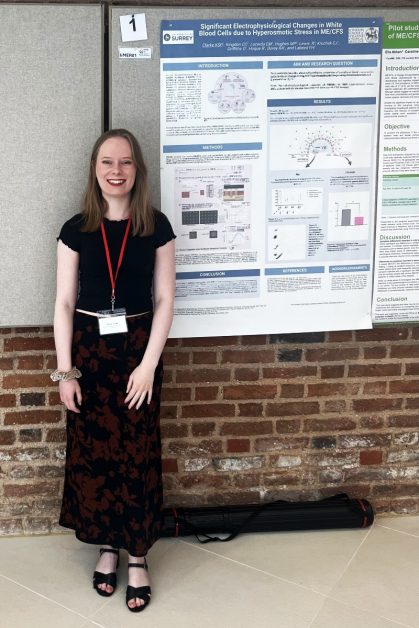
Therefore, the aim of the first chapter of my PhD was to explore the electrical properties of white blood cells in ME/CFS. Furthermore, whether there are any differences which can separate ME/CFS patients, healthy controls, and patients with multiple sclerosis – to act as a much-needed diagnostic biomarker. White blood cells from each group of donors were obtained from the UK ME/CFS Biobank and put in a salty solution for 1.5-hours. I then used two devices to measure the electrical properties (3DEP device – DEPtech, UK) and surface charge (zetasizer – Malvern Panalytical, UK) of the white blood cells. I found the change in the electrical properties of white blood cells before and after salt treatment significantly separated ME/CFS donors from healthy controls and multiple sclerosis donors, with relation to the ionic content in the cytoplasm, ion transport through and across the surface of the cell, and the cell surface charge. These differences are very exciting and show promise towards an adoptable diagnostic biomarker for ME/CFS.
Current Research Focus
After my PhD, I am continuing to investigate these findings as a Research Fellow at the University of Surrey with the research team. I am very thankful to the ME Association and ME Research UK for funding this project, and to all the people who donate samples to the UK ME/CFS Biobank. Data gained from experiments using these samples has been invaluable.
We have come up with a way that separates ME/CFS donors from both healthy controls and multiple sclerosis disease controls using two relatively low-cost and commercially available devices, demonstrating promise towards identifying an adoptable diagnostic biomarker. The main research focus at the moment is to continue data collection to increase the validity of our findings and determine the sensitivity and specificity of the electrophysiological changes as a diagnostic biomarker in a larger donor cohort.
By Dr Krista Clarke
Further information
- The ME Association: Professor Robert Dorey and colleagues update on investigating the electrical properties of blood from people with ME/CFS | July 2, 2024
- The ME Association and ME Research UK announce funding for a study that aims to create a diagnostic test for ME/CFS | October 30, 2023
References
- Broche LM, Bhadal N, Lewis MP, Porter S, Hughes MP, Labeed FH. Early detection of oral cancer – Is dielectrophoresis the answer? Oral Oncology. 2007;43(2):199-203. ↩︎
- Mulhall HJ, Labeed FH, Kazmi B, Costea DE, Hughes MP, Lewis MP. Cancer, pre-cancer and normal oral cells distinguished by dielectrophoresis. Analytical and Bioanalytical Chemistry. 2011;401(8):2455-63. ↩︎
- Liang X, Graham KA, Johannessen AC, Costea DE, Labeed FH. Human oral cancer cells with increasing tumorigenic abilities exhibit higher effective membrane capacitance. Integrative Biology. 2014;6(5):545-54. ↩︎
- Graham KA, Mulhall HJ, Labeed FH, Lewis MP, Hoettges KF, Kalavrezos N, et al. A dielectrophoretic method of discrimination between normal oral epithelium, and oral and oropharyngeal cancer in a clinical setting. Analyst. 2015;14(15):5198-524. ↩︎
- Hughes MP. The surface conductance of red blood cells and platelets is modulated by the cell membrane potential. Electrophoresis. 2023;44(9-10):845-53. ↩︎
- Hoque R, Mostafid H, Hughes MP. Rapid, low-cost dielectrophoretic diagnosis of bladder cancer in a clinical setting. IEEE Journal of Translational Engineering in Health and Medicine. 2020;8:1-5. ↩︎